
Age-related macular degeneration (AMD) is a chronic, usually painless eye condition that damages the macula, the part of the retina responsible for detailed central vision. It can make reading, recognising faces, and seeing fine detail more difficult, while side vision may remain. Early and intermediate AMD may cause no noticeable symptoms, so an eye examination is needed to assess the macula.
For people in Australia, new distortion, sudden reduced central vision or sudden vision loss needs urgent medical assessment. These changes may be caused by wet AMD or another eye emergency and should not be treated as a routine booking matter.
New reduced central vision, new distortion or straight lines appearing wavy, a sudden dark or blurred central patch, or sudden vision loss may be caused by wet AMD or another eye emergency. Seek urgent medical assessment. For sudden or severe vision loss, go to the nearest emergency department or call triple zero (000) if an ambulance is needed.
What is macular degeneration?
The retina is the light-sensitive layer at the back of the eye. At its centre is the macula, a small area that provides the sharp vision needed for tasks such as reading, recognising a face, and seeing fine detail. Macular degeneration affects this central area.
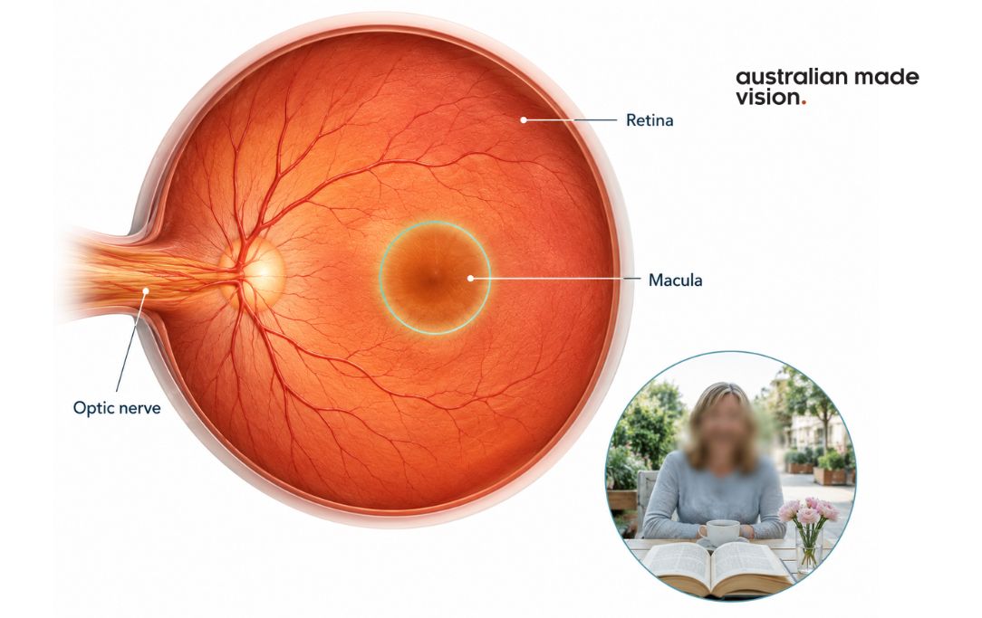

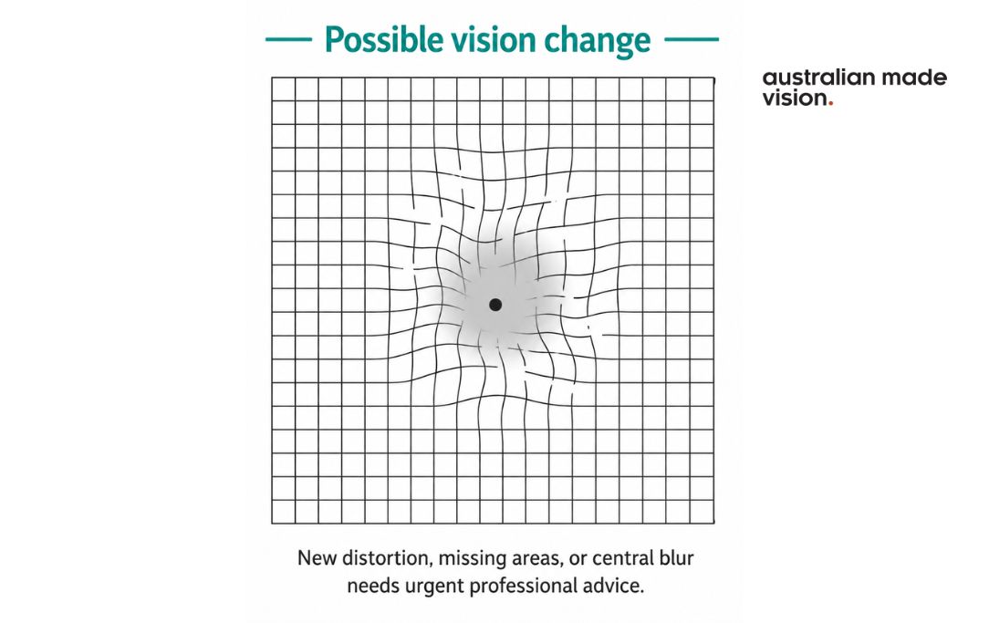
Visual simulation only. Central vision changes and their severity can vary, and this interaction cannot diagnose macular degeneration.
AMD does not usually remove all vision. Peripheral, or side vision is generally preserved, although advanced central vision loss can still have a major effect on everyday activities. The condition may progress slowly in some people and more quickly in others. One eye may also be at a different stage from the other.
Blurred or distorted central vision does not automatically mean a person has AMD. Cataract, refractive change,s and other retinal or neurological conditions can cause similar symptoms. An optometrist or ophthalmologist needs to examine the eyes to identify the cause.
Find Eye Care That Suits Your Needs
Search for an independent optometrist near you and find a local practice that suits your specific eye care needs.
Find an OptometristWhat are the stages and types of AMD?
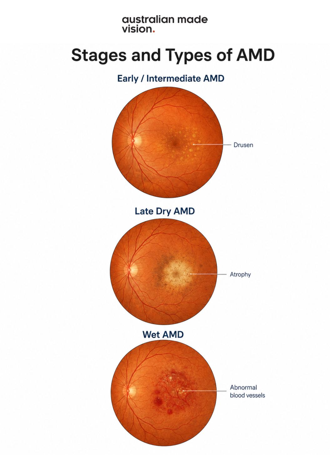
AMD is classified as early, intermediate or late. The stage is based on changes seen during an eye examination rather than symptoms alone.
Early and intermediate AMD
Early and intermediate AMD can involve deposits called drusen under the retina, along with changes in the retinal pigment layer. People with early AMD often notice little or no change in vision. Intermediate AMD may cause some central vision changes, but progression to late AMD is not inevitable.
Late dry AMD or geographic atrophy
Late dry AMD is also called geographic atrophy. It develops when retinal cells gradually deteriorate, creating areas where the retina no longer works normally. The resulting central vision loss is usually gradual, although the rate and pattern differ between people.
Wet or neovascular AMD
Wet AMD occurs when abnormal blood vessels grow beneath the retina and leak fluid or blood. Central vision changes can be sudden and severe. Straight lines may appear wavy or bent, or a new blurred, dark or empty patch may develop near the centre of vision.
Wet AMD is an urgent condition because delays in assessment and treatment can increase the chance of permanent central vision loss.
Dry and wet AMD can occur in the same eye, and a person can have different stages in each eye.
Find Eye Care That Suits Your Needs
Search for an independent optometrist near you and find a local practice that suits your specific eye care needs.
Find an OptometristWhat signs and symptoms can affect central vision?
AMD may be present before a person notices a change. As the condition progresses, symptoms can include:
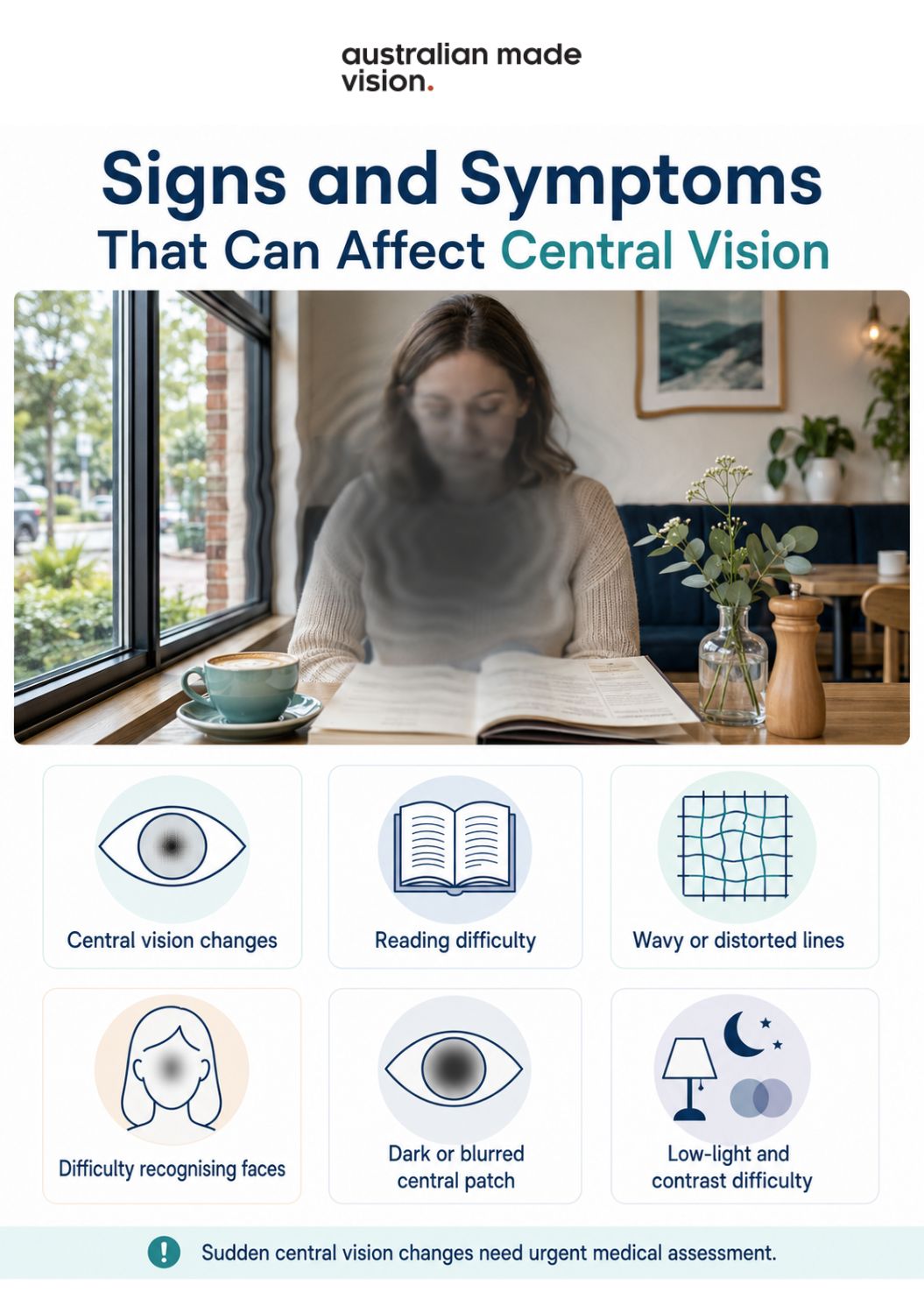
- gradual or sudden worsening of central vision
- difficulty reading or seeing fine detail, even with glasses
- straight lines appearing wavy, bent or distorted
- difficulty recognising faces clearly
- a dark, empty or blurred patch in the centre of vision
- needing brighter light for reading or close work
- reduced contrast or more difficulty seeing in low light
Symptoms can have other causes
These symptoms are not specific to macular degeneration. Persistent blurred or distorted central vision should be assessed, and sudden changes require urgent care. Do not wait for pain: AMD is usually painless.
Is Your Eye
Health on Track?
Complete our free 18-point checklist to check your eye health, spot any gaps, and take charge of your vision.
Start Free Checklist Test →Who may be at higher risk of AMD?
The chance of developing AMD rises with age. A family history of AMD and smoking are also established risk factors. Having one or more risk factors does not mean that AMD will develop, and people without a known family history can still be affected.
Australian eye-health guidance also supports general measures such as not smoking, regular physical activity and a balanced diet. These measures may support overall eye and cardiovascular health, but they do not guarantee that AMD will be prevented or stopped.
An eye-care professional can discuss individual risk and whether the macula should be monitored more closely.
How is macular degeneration assessed in Australia?
An optometrist or ophthalmologist can check vision and examine the retina and macula. The assessment may include reading a vision chart, looking at the back of the eye, and comparing each eye separately. Pupil-dilating drops may be used when a wider view of the retina is needed.
Depending on the clinical findings and the services available, other tests may include:
- Retinal photography, which records an image of the retina for assessment and future comparison.
- Optical coherence tomography (OCT), a scan that creates detailed cross-sectional images of the retinal layers and can show fluid or structural change.
- Angiography, a specialist test that uses dye and imaging to examine retinal blood vessels when needed.
The roles of optometrists and ophthalmologists
An optometrist may detect signs of AMD, monitor changes and arrange referral when specialist assessment is needed. An ophthalmologist is a medical eye specialist who diagnoses and manages treatment for conditions such as wet AMD and geographic atrophy. The exact tests, equipment, fees and referral process vary between practices and services across Australia.
What an Amsler grid can and cannot do
An Amsler grid is a square grid with a dot in the centre. It can help someone at risk of AMD or already diagnosed with AMD notice a change in central vision between eye examinations. The grid should be viewed one eye at a time, using normal reading glasses if required, at the usual reading distance and in good light.
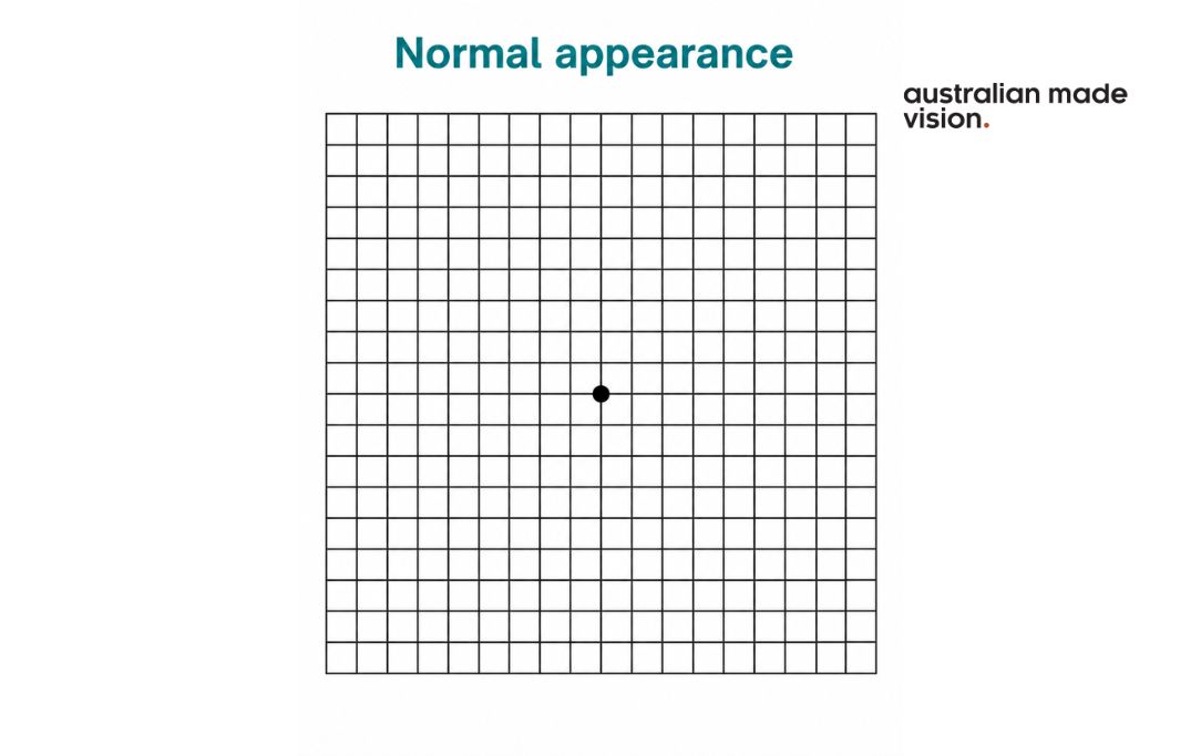
A new wavy, broken or missing area on the grid needs urgent professional advice. The grid cannot diagnose AMD and does not replace an eye examination. Follow the monitoring schedule recommended by the treating eye-care professional rather than relying on a general timetable.
Find Eye Care That Suits Your Needs
Search for an independent optometrist near you and find a local practice that suits your specific eye care needs.
Find an OptometristHow may AMD be monitored, managed, or treated?
AMD management depends on the stage of the condition, the examination findings and the person’s general health. There is no single approach that suits everyone, and treatment decisions should be made with an optometrist, ophthalmologist or other appropriate health professional.
Monitoring and general health measures
Early or intermediate AMD may be monitored through regular examinations and retinal imaging when clinically appropriate. Not smoking is an important modifiable step. Australian guidance also supports regular exercise and a varied diet containing vegetables, fruit and fish as part of general health care.
These measures cannot reverse retinal damage and should not be presented as a substitute for assessment or treatment.
AREDS2 supplements
AREDS2 supplements may be discussed for selected people with intermediate AMD, or late AMD in one eye only. They are not a cure, cannot restore vision already lost and are not recommended for everyone with AMD. They are generally not used to prevent AMD in people without the condition.
Vitamins and minerals can interact with medicines or be unsuitable for some health conditions. Ask an eye-care professional and a GP or pharmacist whether an AREDS2 product is appropriate before starting one.
Treatment for wet AMD
Wet AMD is commonly treated by an ophthalmologist with medicines that block vascular endothelial growth factor, known as anti-VEGF medicines. These medicines are delivered by injection into the eye after local anaesthetic and antiseptic preparation. Treatment usually requires ongoing monitoring, and the injection schedule depends on how the eye responds.
Anti-VEGF treatment can reduce leakage from abnormal blood vessels and may stabilise or improve vision for some people. It does not cure AMD, and treatment benefits and risks vary. Sudden pain, worsening redness or an unusual vision change after an injection should be reported promptly to the treating service.
Treatment discussions for geographic atrophy
Macular Disease Foundation Australia reports that two treatments are registered by the Therapeutic Goods Administration for geographic atrophy. These treatments are not suitable for every person with late dry AMD. Eligibility, expected benefit, treatment burden, risks, availability and costs need to be discussed with an ophthalmologist using current Australian information.
Because treatment options and access can change, this section should be checked again immediately before publication and during clinical review.
Can glasses treat macular degeneration?
Glasses can correct short-sightedness, long-sightedness, astigmatism or other refractive blur that occurs alongside AMD. They cannot repair damage to the macula. Some people may benefit from low-vision aids, stronger task lighting, magnification or assistive technology to make better use of their remaining vision.
Living with AMD and finding support in Australia
Central vision changes can make detailed tasks harder, but practical support may help. A low-vision assessment can look at the tasks that matter to the individual and consider options such as magnifiers, improved lighting, contrast changes, electronic readers, screen enlargement and other assistive technology.
Macular Disease Foundation Australia provides information, support and a national helpline. Vision Australia also offers low-vision and blindness services. Eligibility, costs and available services vary, so contact the organisation or treating service for current details. Family members and carers can also be included in discussions about home adjustments, transport and communication preferences when the person wishes.
When should you seek routine, prompt or urgent care?
The appropriate next step depends on how quickly the change started and how severe it is.
- Urgent or emergency assessment: sudden vision loss, new reduced central vision, new distortio,n or a sudden dark or blurred central patch. Seek emergency medical advice or attend an emergency department. Call triple zero (000) if an ambulance is needed.
- Prompt eye assessment: persistent or unexplained central blur, distortion, difficulty seeing faces or a noticeable change in detailed vision.
- Routine discussion: family history of AMD, questions about monitoring, an existing AMD diagnosis, or whether supplements may be appropriate.
Emergency symptoms should not be directed to a routine online booking pathway. For non-urgent concerns, an eye examination can help identify the cause and the next step.
Questions to ask an optometrist or practice
- Will the examination include a check of the macula?
- Is OCT or retinal photography available, and could extra fees apply?
- What happens if wet AMD is suspected?
- Who will monitor the condition, and how are referrals handled?
- Could AREDS2 be appropriate for this stage of AMD?
- How should an Amsler grid be used, and what change needs urgent action?
- Which low-vision or support services may be suitable?
A measured next step
Sudden central vision loss or new distortion needs urgent medical assessment. For persistent but non-urgent changes, family-history questions or AMD monitoring, an eye examination can help determine what is happening.
Australian Made Vision publishes general eye-health information and provides a directory of participating independent practices. Use the directory to find an independent optometrist in Australia, then confirm services, fees, and availability directly with the practice. Appointments, examinations, referrals and treatment are provided by the individual practice and clinicians.
Find Eye Care That Suits Your Needs
Search for an independent optometrist near you and find a local practice that suits your specific eye care needs.
Find an OptometristMacular Degeneration FAQs
Can macular degeneration cause total blindness?
AMD mainly affects central vision and usually leaves peripheral vision. It does not generally cause complete blackness by itself, but advanced AMD can cause severe vision impairment and make detailed tasks very difficult.
Is macular degeneration hereditary?
Family history increases risk, but AMD is not determined by one factor alone. A parent or sibling with AMD is a reason to discuss personal risk and examination timing with an eye-care professional, not proof that the condition will develop.
How quickly can wet macular degeneration affect vision?
Wet AMD can cause rapid changes in central vision. New distortion, reduced central vision, or a sudden dark or blurred area requires urgent assessment rather than waiting for a routine appointment.
Can glasses correct vision loss from macular degeneration?
Glasses may correct a separate prescription problem, but they cannot repair the damaged macula. Low-vision aids and changes to lighting, magnification and technology may help with particular tasks.
Are AREDS2 supplements suitable for everyone with AMD?
No. Their possible benefit depends on the stage of AMD and which eye is affected. They are not a cure and may be unsuitable because of medicines or other health conditions. Professional advice is needed before use.
When is sudden distortion or central vision loss an emergency?
New wavy or bent lines, new reduced central vision, a sudden central dark, or blurred patch, or sudden vision loss needs urgent medical assessment. Sudden or severe vision loss should be treated as an emergency.
Australian Made Vision is an independent directory that helps patients find optometrists across Australia who may offer Australian-made spectacle lenses. We do not provide clinical services and recommend contacting practices directly to confirm the products and services they offer.
- Healthdirect Australia – Macular degeneration
- Macular Disease Foundation Australia – AMD overview</